
DNA replication in Bacteria
Spying the dynamics inside the replisome
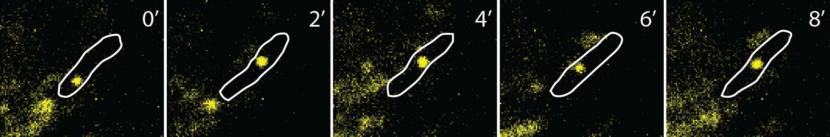
Outline of E.coli with a fluorescent focus representing a single molecule of mMaple fused to the DnaB helicase. Pictures taken every 2 minutes.
The replisome is composed of multiple different proteins, some of them present in more than one copy per replication fork. Once assembled, the bacterial replisome is able to copy millions of bases pairs. In contrast with this stability, cycles of binding and unbinding of individual subunits define how DNA replication is regulated at initiation, how it achieves periodic cycles of synthesis at the lagging strand, how does it prevent premature termination, how the replisome bypasses lesions on DNA, and how does replication terminates. There are still many gaps in our understanding on the dynamics inside the replisome. Our ignorance is partly due to the difficulty of determining the fate of individual components of the replisome while actively synthesising DNA.
By focusing sensitive microscopes on cells undergoing DNA replication, we use quantitative approaches to determine how often does the replisome replace its components during DNA replication. This information will allow us to understand how DNA replication is carried out and what factors determine the stability of replisome as a protein assembly. We are also interested in observing the recruitment and unbinding of components at initiation and termination, in order to better understand these processes.
Examples of single copies of replisome subunits in live E.coli. The first one shows molecules of the epsilon subunit of DNA Polymerase III, both immobile and diffusing,captured at rates of 20 ms (50 pic per second). The second shows immobile copies of the DnaB helicase bound for several minutes to the chromosome (one pic every 20 seconds)